
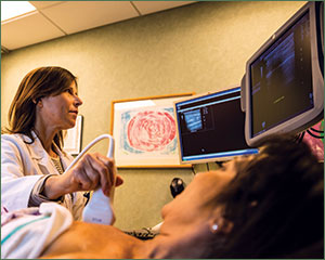
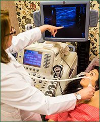
What is a Breast Ultrasound?
Breast ultrasound is a painless examination that uses high-frequency sound waves to obtain images of the inside of the breast. High-resolution technology produces breast images that are extremely clear and show an impressive amount of fine detail.
Images are displayed on a video monitor that both you and the radiologist view, and discuss, together.
Why would I need a Breast Ultrasound?
Montclair Breast Center uses breast ultrasound as both a screening and diagnostic tool. Ultrasound is used as a diagnostic tool when an abnormality is found on physical breast examination, mammography or MRI.
Women with dense breast tissue often receive an ultrasound in addition to a mammogram. Correlating the physical findings from breast examination with the visual images on ultrasound can save women from unnecessary biopsies or help provide an early diagnosis of breast cancer. This represents the most sophisticated refinement of the clinical breast examination. In addition, with an ultrasound examination many mammographic abnormalities can be resolved and surgery can be avoided. Cysts, for example, are easily seen and do not need a biopsy. Complex cysts and solid masses are sampled using minimally invasive needle biopsy techniques.
Correlating the physical findings from breast examination with the visual images on ultrasound can save women from unnecessary biopsies and/or help provide an early diagnosis of breast cancer. With the addition of an ultrasound, many mammographic abnormalities can be resolved and surgery can be avoided. For example, ultrasound’s can show whether a lump found during an exam is a solid mass or a cyst (fluid collection).
If you are pregnant or breastfeeding, an ultrasound is a safe tool for “seeing” inside the breasts and may be recommended.
What is an “Ultrasound Guided Biopsy?”
This minimally invasive technique is used to evaluate abnormalities. A small sample of breast tissue is collected using a special needle biopsy device guided by continuous, high-resolution ultrasound images. We use this technique as our first option when the mammogram abnormality can also (or only) be seen on an ultrasound.
The procedure is quicker and more comfortable than stereotactic biopsy.
There is no discomfort and only local anesthetic is needed. No stitches are necessary. Results are available within 24-48 hours.
What is a “Guided Aspiration of Cysts?”
To confirm an abnormality is definitively a cyst, your radiologist may remove the cyst contents for evaluation.
This is a fast and painless procedure. Cyst contents generally do not need to be sent for analysis.


